Keratoconus and corneal cross-linking
Keratoconus is a condition in which the cornea, the clear front window of the eye, gradually thins and bulges into a cone shape. It usually starts in the teenage years or twenties and can progress for years. It causes distorted, blurry vision that glasses cannot always correct.
Signs to look for
- Vision that keeps changing, with frequent new glasses prescriptions
- Increasing astigmatism or short-sightedness
- Ghosting, streaking or glare, particularly at night
- Frequent eye rubbing, which is linked to keratoconus and is best avoided
- Contact lenses that no longer fit or feel comfortable
People with allergies, eczema or a family history of keratoconus have a higher chance of developing it. Keratoconus is also a reason to be cautious about laser vision correction, which is why detailed corneal scans matter before any laser procedure.
How it is diagnosed
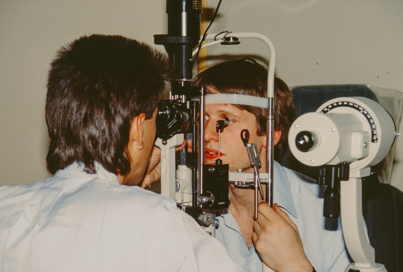
Diagnosis uses corneal mapping (topography or tomography), which measures the shape and thickness of the cornea in fine detail. These scans can detect keratoconus before it is obvious on a standard eye test. Repeat scans over time show whether it is progressing.
Corneal cross-linking (CXL)
Cross-linking is a treatment designed to stop keratoconus from getting worse by strengthening the cornea. Riboflavin (vitamin B2) drops are applied to the cornea, which is then exposed to a controlled dose of ultraviolet A light. This creates new bonds between the collagen fibres, making the cornea stiffer.
- It aims to halt progression. It does not usually restore normal vision, though some people see modest improvement.
- It is most effective when done early, while vision is still good, and when progression has been shown.
- In the standard (“epi-off”) technique the surface layer of the cornea is removed so the drops can soak in. It is the most established approach. Recovery involves several days of discomfort and slow visual recovery.
- Variations, including techniques that leave the surface layer in place (“epi-on”) and accelerated protocols, are being studied. Availability in Australia can differ, and their effectiveness compared with the standard approach is still being assessed. Dr Abell offers epithelium-on accelerated, oxygen-assisted corneal collagen cross-linking.
After cross-linking
Once keratoconus is stable, glasses or specialist contact lenses (such as scleral lenses) can often restore good functional vision. Other treatments, such as corneal ring segments, may help in selected cases. A small number of people with advanced disease or scarring eventually need a corneal transplant. See our article on corneal transplant techniques.
Risks of cross-linking
Risks include infection, corneal haze or scarring, delayed healing, discomfort, and, in some cases, continued progression despite treatment.
When to see a specialist
See an optometrist or ophthalmologist if your vision is changing, your prescription keeps changing, or your optometrist has spotted an irregular cornea. Early referral allows treatment before significant vision is lost.
This article is general information and is not a substitute for individual medical advice. All surgical and invasive procedures carry risks.